

Text written in Basque and translated automatically by Elia without any subsequent editing. SEE ORIGINAL
Find out in 3D how SARS-CoV-2 replicates inside cells
2023/11/20 Galarraga Aiestaran, Ana - Elhuyar Zientzia Iturria: Elhuyar aldizkaria
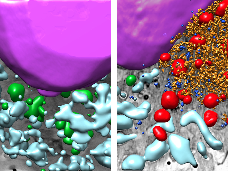
An uninfected cell on the left; infected on the right. Purple core, green healthy mitochondria, red damaged mitochondria, light blue vacuoles, yellow virus machinery and dark blue viral particles. When the virus that produces COVID-19 infects lung cells, they've seen how intracellular structures change in three dimensions by combining three techniques of molecular biology, virology and microscopy, including cryocellular tomography with soft X-rays.
The work has been led by CNB-CSIC researchers and the results have been published in ACS Nano. It is explained that, comparing an infected cell with the non-infected one, it becomes evident that the virus replication machine produces vesicles and tubules, and causes stress in organelles, such as mitochondria and endoplasmic reticulum.
These images allow researchers to better understand how the virus replicates and how therapies work.

eu es fr en ca gl
Gai honi buruzko eduki gehiago
Elhuyarrek garatutako teknologia

.jpg)